
Немногие ценят зрение, пока оно у них есть, и мало кто знает, что глаза — зеркало не только души, но и здоровья. Сегодня с помощью современного оборудования в специализированной клинике, даже не прикасаясь к телу человека, по состоянию его глаз можно получить представление об общем состоянии организма.
Текст: PETER TOKARSKI, CULT OF VISION
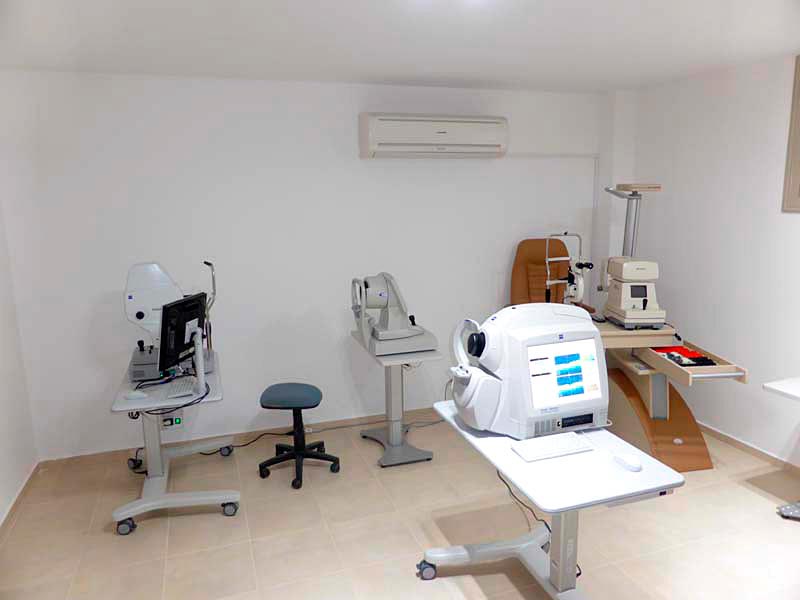
В Cult of Vision мы проводим подобную диагностику на ультрасовременном оборудовании и можем определить состояние не только ваших глаз, но и здоровья в целом.
ОКТ исследование: 3D томография глаза
Это самый инновационный и глубокий вид исследования, который позволяет провести сканирование передней и задней части глазного яблока.
Такое сканирование дает оптометристу детальное изображение сетчатки, по которому можно определять, отслеживать и контролировать любые изменения. На сегодняшний день это единственная процедура, которая позволяет увидеть внутренние структуры глаз. Исследуются также и снимки роговицы. Анализируя их, можно на ранних стадиях диагностировать любые отклонения от нормы. Все остальные виды диагностики позволяют увидеть лишь поверхность этих структур.
ОКТ выявляет ранние симптомы макулярной дистрофии, глаукомы, отслоения сетчатки, кератоконуса, дистрофии роговицы и других заболеваний глаз. Процедура занимает всего несколько минут и не приносит дискомфорта, так как не подразумевает прямого контакта оборудования с глазами.
Фотографии сетчатки
Используя немедиатрическую фундус-камеру последнего поколения, мы можем провести очень тщательные исследования желтого пятна, фовеолярной зоны, сетчатки и диска зрительного нерва. Эти исследования дают возможность нашим специалистам вовремя диагностировать серьезные заболевания, такие как глаукома и возрастная дегенерация макулы. Фотографии сетчатки также помогают диагностировать диабет и нарушения деятельности сердечно-сосудистой системы (эндокардит и гипертензию).
Топография роговицы
Этот вид исследования глаза дает оптометристу информацию о нарушениях в роговой оболочке. Специалист проверяет поверхность роговицы и получает подробные данные об астигматизме и кератоконусе. Это также позволяет создать индивидуальные контактные линзы. Опытные специалисты благодаря современным приборам гарантируют самые лучшие результаты при индивидуальной коррекции зрения.
Осмотр глаза с помощью щелевой лампы
Оптометрист проводит осмотр передней и задней части глаза с помощью микроскопа щелевой лампы. Это позволяет специалисту исследовать веко, роговицу, радужную оболочку, хрусталик глаза и видимую часть сетчатки и дать пациенту подробную информацию о состоянии здоровья глаз.
Зрение, пожалуй, самое важное из пяти чувств, и не стоит им пренебрегать. Некоторые из упомянутых заболеваний невозможно диагностировать без специального оборудования. Обращайтесь к опытным специалистам, в распоряжении которых самая современная техника, и помните, что ранняя диагностика — это половина успеха лечения!
Адрес: Corner of 25 Martiou and Kostaki Pentalioti, Solomou Square., Пафос, 26955955


